
 1800 867 1390
In this webinar and Q&A, solicitor and former regulator David Gardner provides exclusive insights...


100% online
$1495
82 hrs
 |
 |
 |


Attending Physician, Dermatology Service, Memorial Sloan Kettering Skin Cancer Center, New York, USA
Professor Ashfaq A. Marghoob is a board-certified dermatologist specialising in the diagnosis and treatment of cancers of the skin. He is the director of Memorial Sloan Kettering’s regional skin cancer clinic in Long Island and consults and treats patients in the centre’s outpatient facility in Manhattan.
Although providing the best care possible for his patients remains his primary goal, Ashfaq also remains committed to education and clinical research, with the hope of educating physicians and the public about the importance of early skin cancer detection to save lives.
He is active in clinical research and has published numerous papers on topics related to skin cancer with an emphasis on melanoma, atypical/dysplastic nevi, and congenital melanocytic nevi. Ashfaq’s research interests are focused on the use of imaging instruments such as photography, dermoscopy, and confocal laser microscopy to recognise skin cancer early in its development.

Professor and Course Coordinator MMed (Skin Cancer) Program School of Medicine, The University of Queensland
Professor Cliff Rosendahl currently works in Brisbane as a primary care practitioner with a special interest in skin cancer. He also has an interest in research as the clinical developer and Director of the Skin Cancer Audit Research Database (SCARD). His other main area of research has been in evaluating dermatoscopic clues for the diagnosis of both pigmented and non-pigmented skin malignancy in collaboration with colleagues at The University of Queensland, Australia and the Medical University of Vienna, Austria.


Associate Professor Skin Cancer Medicine, The University of NSW
Associate Professor John Pyne was conferred with a Master of Medicine at The University of Queensland, then held a position of Program Director for The University of Queensland Skin Cancer Masters from 2007 until 2013. The thesis for John’s PhD was on the correlations between dermatoscopy and histopathology for BCC and SCC. He is also a past President and Fellow of the Skin Cancer College of Australasia.
John has a main interest in the optical imaging of early primary non-lymphoid cutaneous malignancy and correlations with histopathology. He is currently investigating surgical margins in management of these cancers. John has teaching appointments at The Universities of Queensland, New South Wales and Western Sydney.

Honorary Lecturer in Pathology, The University of Queensland
Dr Simon Clark runs the pathology component of the postgraduate certificate courses in skin cancer medicine at The University of Queensland and lectures in the Masters of Medicine course. He has been involved in dermatopathology education for more than 20 years, training registrars in dermatology, pathology and plastic surgery. More recently he has been active in GP education. One of the best known dermatopathologists in Australia, Simon was recently appointed a visiting professor in dermatology at the Tehran University of Medical Sciences.

Dermatologist, Santa Maria Nuova Hospital, Reggio Emilia, Italy
Dr Elvira Moscarella is a dermatologist at the Santa Maria Nuova Hospital in Reggio Emilia, Italy. She acquired her medical degree in 2005 at the Second University of Naples before completing her residency in dermatology and venereology at the University’s Department of Dermatology. In 2008, Elvira undertook further education in dermoscopy and confocal microscopy. She is a member of the European Academy of Dermatology and Venereology and the International Dermoscopy Society, and is Editor in Chief of the latter’s newsletter and case of the month. Elvira’s main interests are in dermoscopy and reflectance confocal microscopy, and their use in skin cancer medicine.


In this webinar and Q&A, solicitor and former regulator David Gardner provides exclusive insights...
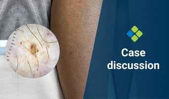
This week's case discussion, submitted by Dr Heather Lawson, features a 64-year-old female patient...

Mood changes in the postpartum period are extremely common, ranging from mild and transient “baby...


