Recognising risk beyond Fitzpatrick I-III: What GPs need to know about dermoscopic diversity and...
.jpg?height=200&name=HCE%20HubSpot%20blog%20images%20600x350%20(27).jpg)
Learn the foundations of primary care skin screening.
Designed for Registered Nurses, Nurse Practitioners, and allied health professionals working in specialist skin clinics.
%20(4).webp)
Learn the foundations of primary care melanography, from identifying common types of benign and malignant skin lesions through to best practice approaches for skin cancer screening and reaching a confident diagnosis.
- Develop the skills to play a vital role in the early detection and management of melanoma, thereby helping to improve patient outcomes by ensuring timely intervention and comprehensive care.
- Ideal foundation course for nurses and allied health professionals with little knowledge in lesion screening.
- Presented by world-leading experts in dermoscopy.
- Reviewed by the International Dermoscopy Society, and Registered Nurses have provided documentation on Scope of Practice for nurses to facilitate their CPD in this field.
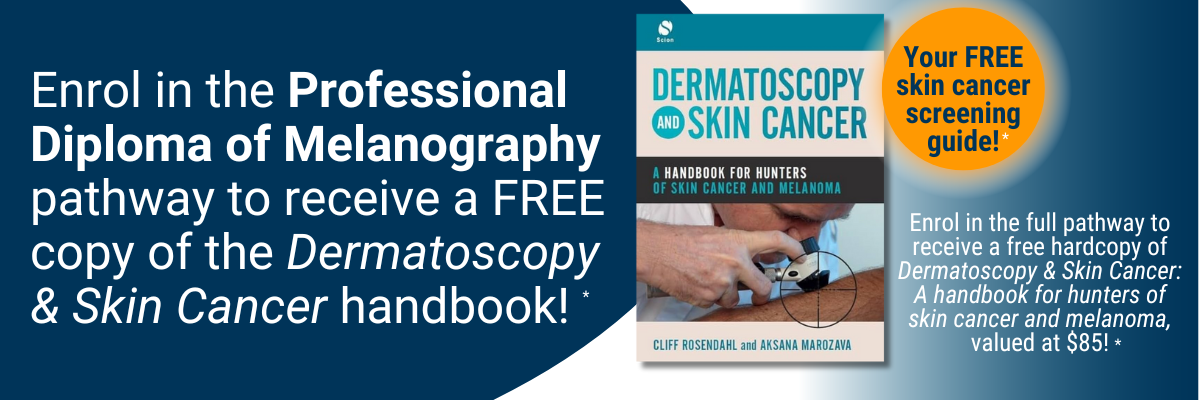
* T&Cs apply: Stocks are limited; offer only available while stocks last and only available to those who enrol in, and pay for, the full three-stage Melanography pathway bundle. Books posted within Australia only; you must have a valid Australian postal address to receive the book. The book will be automatically mailed to you upon successful enrolment.
- Accurately screen and differentiate skin lesions for referral to a doctor for diagnosis and treatment.
- Learn to assess commonly encountered skin malignancies, including melanoma, BCC and SCC.
- Identify pigmented and non-pigmented skin lesions.
- Confidently identify benign and malignant non-melanocytic lesions and pink lesions in your patients.
- Understand the Chaos & Clues method for simple lesion assessment and faster diagnoses.
- Provide early intervention for your patients.
Get unlimited access to all course content, additional learning materials, ongoing post-course support, and more.
What is a melanographer?
A melanographer is a nurse or health professional whose primary role is to conduct skin assessments and identify suspicious skin lesions for referral to a doctor.
1. Skin assessments and diagnostic procedures: Melanographers are skilled in conducting comprehensive skin assessments to identify suspicious lesions that may indicate skin cancer, using a combination of tools including a dermatoscope and taking images. Melanographers may assist doctors during diagnostic procedures like biopsies.
2. Patient education: Melanographers educate patients about skin cancer prevention and early detection, sun protection, self-examination, and skin checks.
3. A multidisciplinary approach: Melanographers work within a multidisciplinary team which may include GPs, dermatologists, oncologists and other healthcare professionals involved in caring for skin cancer patients.
This module focuses on the foundations relating to skin cancer medicine. Participants will acquire the knowledge required to safely and confidently diagnose and treat commonly encountered skin lesions. It is the ideal starting point to build core knowledge in skin cancer management and acquire vital diagnostic skills and basic management techniques to provide effective care to patients.
Unit one introduces the foundation information required for skin cancer medicine and outlines the content of the course. Unit two describes the BEST protocol (BEnign or Suspicious Test) and divides lesions into benign or suspicious with guidelines and clinical images of pigmented skin lesions. Benign lesions require no action and suspicious lesions require biopsy or referral. A quiz with clinical and dermoscopic images to select benign or suspicious lesions is provided, with contextual answers as part of this unit.
Unit three discusses the importance of skin checks and commences with a table dividing assessment of skin cancer risk into three categories: high, medium and low risk and includes patient self-examination and opportunistic skin check. The unit describes how to perform a skin examination and includes lesion history, general medical history, allergies and current medications.
The unit includes a video of a systemic total skin examination. Unit four describes partial and complete punch and shave biopsy techniques.
The unit concludes with a patient case showing cylindrical portion of skin bearing a dark pigmented surface for which biopsy is recommended. The pathology report is provided as the lesion was partially excised and sent for examination.
This module focuses on the big three – BCC, SCC and melanoma relating to skin cancer medicine. Participants will learn about the clinical and dermoscopic features of BCCs, biopsy options, pathology and treatment approaches. The module then moves to solar keratosis and examines the use of topical non-surgical treatments.
Unit two continues with field-directed treatment methods including photodynamic therapy, and lists the pros and cons of available treatments. Two patient cases are included.
Unit three covers SCC lesions and outlines the clinical and dermoscopic features, biopsy options, pathology and treatment approaches. Clinical cases including images are provided for various SCCs.
Unit four focuses on melanomas and sentinel lymph nodes and again outlines the clinical and dermoscopic features, biopsy options, pathology and treatment approaches. Clinical cases are provided for various melanomas.
This module focuses on the approach to pigmented skin lesions relating to skin cancer medicine. Participants will acquire the knowledge required to safely and confidently diagnose and treat commonly encountered skin lesions. It is the ideal starting point to build core knowledge in skin cancer management and acquire vital diagnostic skills and basic management techniques to provide effective care to patients.
Unit one discusses common benign lesions including freckles, solar lentigo, seborrheic keratosis, haemangioma, dermatofibroma and blue naevi. Normal and dermoscopic clinical images are provided to demonstrate patterns and assists with analysis. Benign lesions can mimic skin cancers and careful examination is required to decide management steps. The signs of common benign lesions that may be skin cancer are discussed in detail. A medico-legal case is included for a patient where a melanoma was masquerading as a benign lesion.
Unit two begins with the description of dermoscopy including examples of dermoscopic devices used for this technique. The 3-point checklist consists of asymmetry in colour or structures, atypical network and blue-white structures (white scar-like depigmentation or blue pepper-like, globular or structure-less areas), showing various clinical dermoscopic images of lesions.
This unit covers dysplastic naevi and melanoma. A table is shown of the relationship between nevus, dysplastic nevus and melanoma and includes a table of relative risk factors for melanoma. Several types of melanomas and treatment options are listed and supported by clinical images. The unit concludes with key points for detecting benign or suspicious lesions.
This module focuses on the approach to non-pigmented skin lesions relating to skin cancer medicine. Participants will acquire the knowledge required to safely and confidently diagnose and treat commonly encountered skin lesions. It is the ideal starting point to build core knowledge in skin cancer management and acquire vital diagnostic skills and basic management techniques to provide effective care to patients.
This unit starts with a clinical description of solar keratosis and outlines treatment options including field treatment. Attention is also paid to Bowen’s disease (a very early form of skin cancer), including symptoms, diagnosis (punch biopsy) and treatment. Dermoscopic images of both conditions are provided.
This unit describes keratocanthoma, a low-grade malignancy that requires a deep incision biopsy, and also discusses squamous cell carcinoma, a common cancer due to chronic, cumulative, sun exposure. Treatment options include surgery, curettage and cautery, specialist referral or radiation therapy. Clinical images of both conditions are provided.
Unit three describes basal cell carcinoma, the most common human cancer due to intermittent excessive sun exposure. BCC can be classified as non-aggressive (superficial, nodular) or aggressive (sclerosing / infiltrating / micronodular / multifocal / solid). Clinical images of the different types of basal cell carcinoma, diagnosis and treatment options are included.
This module introduces the Chaos and Clues diagnostic method for identifying whether a skin lesion is benign or suspicious. This algorithm is based on revising pattern analysis to identify chaos, clues and exceptions, to assist with the diagnosis and management of skin lesions. Extensive images and examples are provided. In this module, the Chaos and Clues method is used to assess pigmented skin lesions such as melanomas, pigmented basal cell carcinomas (BCCs) and pigmented squamous cell carcinoma (SCCs). The module also considers the four exceptions when considering a biopsy.
This module identifies the four most common types of benign non-melanocytic skin lesions commonly seen in daily practice. It explains in detail the use of dermoscopy in diagnosing seborrheic keratosis, dermatofibroma, vascular tumours and sebaceous hyperplasia including numerous dermoscopic images. The module explains the factors influencing the prevalence and morphology of these benign lesions. The module concludes with three management rules to follow in order not to miss melanoma that mimics a benign non-melanocytic tumour.
This module focuses on malignant non-melanocytic skin lesions commonly seen in daily practice. It explains in detail the use of dermoscopy in diagnosing keratinocyte carcinomas, basal cell carcinoma and squamous cell carcinoma. Dermoscopic images and pattern analysis are used throughout the presentation to differentiate between benign and malignant skin lesions.
This module focuses on the general aspects of the dermoscopic examination of pink tumours. It explains the patterns associated with melanocytic and non-melanocytic skins tumours in detail. This module also explains the contact and non-contact dermoscopy methods used when examining skin tumours. Dermoscopic images are used throughout the presentation to identify if the pink tumours are benign or suspicious based on vascular structures, arrangements, and clues. The module concludes with management rules to follow in order not to miss amelanotic melanoma.
Participants will learn about skin imaging techniques for detecting melanoma and non-melanoma skin cancers. Various techniques are outlined commencing with (digital) dermatoscopy, listing physical foundation, indications, evidence and target groups. Total body photography is described and illustrated by images. A summary is given of indications, evidence and target groups. The module looks at reflectance confocal microscopy (RCM), explaining the concept of the method, in which cases it is used and clinical indications. A chart is shown of possible combination of digital dermatoscopy with RCM. A summary is given of indications, evidence and target groups. The course then discusses optical coherence tomography, high frequency ultrasound, electric impedance spectroscopy, multispectral analysis and raman spectroscopy listing physical foundation, indications, evidence and target group.

Professor at the Department of Dermatology, Medical University of Vienna, Austria
Professor Harald Kittler has a special clinical interest in dermoscopy of pigmented skin lesions. His main research interest is digital dermoscopy, follow-up of pigmented skin lesions, and computer assisted digital dermoscopy. Harald has been working for 10 years in the field of dermoscopy and has published a number of scientific articles especially in the field of digital dermoscopy and dermoscopic follow-up of melanocytic nevi.

Professor and Course Coordinator MMed (Skin Cancer) Program School of Medicine, The University of Queensland
Professor Cliff Rosendahl currently works in Brisbane as a primary care practitioner with a special interest in skin cancer. He also has an interest in research as the clinical developer and Director of the Skin Cancer Audit Research Database (SCARD). His other main area of research has been in evaluating dermatoscopic clues for the diagnosis of both pigmented and non-pigmented skin malignancy in collaboration with colleagues at The University of Queensland, Australia and the Medical University of Vienna, Austria.

Professor Philipp Tschandl graduated from the Medical University of Vienna where he also obtained his PhD degree. His main research field is skin cancer, especially its early diagnosis through dermatoscopy, and the teaching of that method. He is an executive board member of the International Dermoscopy Society, secretary of the National Dermatopathology Society in Austria, and member of the Fostering Trainee Education Committee of the EADV.
Philipp is teaching dermatology in the core curricula of Human and Dental Medicine, lectures continuously on dermatoscopy at national and international meetings and workshops, and co-organised the 4th World Congress of Dermoscopy 2015 in Vienna. He has published more than 30 peer-reviewed scientific articles and is co-author of a major dermatoscopy textbook which has been translated into multiple languages.

Chief Medical Officer,
National Skin Cancer Centres
Professor David Wilkinson is a registered general practitioner and public health medicine expert. Prior to taking up the role as Chief Medical Officer with National Skin Cancer Centres, he was Deputy Vice-Chancellor of the Macquarie University, Sydney for eight years, and before that was Dean of Medicine at The University of Queensland for nine years.
Since 2004, David’s clinical work has focused on skin cancer medicine in primary care. He has published research papers on the topic, designed and led development of the only Master of Medicine degree in skin cancer, and helped develop and present a suite of skin cancer short courses delivered by HealthCert. He has taught almost 7,000 GPs the basics of skin cancer medicine in primary care.

Head of the Dermatology Clinic of the University of Trieste, Italy
Associate Professor Iris Zalaudek is a board-certified dermatologist and Head of the Dermatology Clinic of the University of Trieste, Italy. Since 2016, she has been President of the International Dermoscopy Society, and was previously the Research Director of the Non-Melanoma Skin Cancer Unit at the Medical University of Graz, Austria.
Her main research fields are related to dermato-oncology and include non-invasive skin imaging techniques, as well as topical and systemic treatment of skin cancer. Moreover, she is engaged in the development of modern teaching methods such as online distant courses and tele-dermatologic services. She is Director of the Master of Science program entitled "Dermoscopy and Preventive Dermato-Oncology" of the Medical University of Graz, Austria.
Iris has published more than 450 articles, of which 358 (267 full papers) have been cited in PubMed. Her combined publications have received an impact factor of 1003 and a h-index value of 36 (by April 2017). In 2003 her work was awarded by the Hans-Weitgasser Price from the Styrian Association of Dermatologists and in 2008 she was awarded the Best Researcher of the Medical University of Graz, Austria.

Doctor, National Skin Cancer Centres, Berwick
Dr Hamilton Ayres worked in Adelaide as a Plastic Surgery Registrar at Flinders, Repatriation General Hospital and the Royal Adelaide Hospital where his main role was the management of trauma, hand injuries and difficult skin cancers. Hamilton has obtained a Fellowship of the Royal Australian College of General Practitioners and Certificates in Skin Cancer Medicine, Dermatoscopy and Histopathology from HealthCert and The University of Queensland School of Medicine.

$1495
.
Bundle two courses and save 5%, or three courses and save 10% upon enrolment.
Talk to us about deferred payment options, registrar scholarships and special rates.

An excellent course introduction to dermoscopy and relating what you see to the histopathology and applying this in clinical practice. Great lectures and supporting materials.
S. Jan
Excellent! This is a great course that has helped me diagnose many more subtle, early skin cancers especially melanoma in situ. The course was clearly presented, with good pictures and course book. All HealthCert's skin cancer courses have been hugely valuable to my practice!
R. Mundell
I highly recommend this course. I increased my knowledge and developed confidence in using dermoscopy and in diagnosing melanoma and other skin lesions. Every skin lesion I see means so much more now that it has a name. Great involvement from various skin cancer experts and great videos and reference materials.
P. Ishri
I would recommend this course as an important addition to self-directed learning in dermatoscopy. It was a invaluable course to gain the diagnostic tools, knowledge and confidence in managing skin cancer. The course was professionally conducted with excellent presentations.
L. Suntesic
I really enjoyed the level of learning. It is very rewarding to know that I am potentially saving lives. Recently I volunteered with the Lions Cancer Institute for two days, and we screened 158 patients, detected 38 possible melanomas and 83 keratinocyte skin cancers. It was a very successful and rewarding two days, and something I could do confidently because of my learning from this course.
K. Laverty
| RACGP Activity Number | ACRRM Activity Number | Activity Title | Education Hours | Performance Hours | Outcome Hours | ||
|---|---|---|---|---|---|---|---|
| 403783 | 28433 | Acral lesions | 403783 | 28433 | 3.5 | 6 | 0 |
| 403786 | 28434 | Melanoma | 403786 | 28434 | 4.5 | 6 | 0 |
| 403766 | 28428 | The Chaos and Clues method | 403766 | 28428 | 4.5 | 6 | 0 |
| 403764 | 28427 | Algorithms and the elephant approach | 403764 | 28427 | 4 | 6 | 0 |
| 403772 | 28430 | Malignant non-melanocytic lesions commonly seen in the practice | 403772 | 28430 | 4 | 6.5 | 0 |
| 403778 | 28432 | Facial lesions | 403778 | 28432 | 4.5 | 6.5 | 0 |
| 403769 | 28429 | Benign non-melanocytic lesions commonly seen in the practice | 403769 | 28429 | 3.5 | 5.5 | 0 |
| 403775 | 28431 | Melanocytic nevi | 403775 | 28431 | 4 | 5.5 | 0 |
| 800429 | 32973 | Acral Lesions Outcome Improvement Activity | 800429 | 32973 | 0 | 0 | 8.5 |
| Total hours | 32.5 | 48 | 8.5 | ||||
The purpose of outcome measurement activities is to improve your clinical confidence in managing an identified learning gap. Outcome measurement activities are not a requirement of our Professional Certificate of Advanced Certificate courses; they are a requirement for Australian CPD purposes.
HealthCert Education provides a variety of outcome measurements activities to suit your needs:
HealthCert Education offers courses suitable for allied health professionals and all nurses, including Enrolled Nurses, Registered Nurses, and Nurse Practitioners. Please read the course description carefully to select a course that is right for your nursing role.
The Professional Certificate of Melanography is suitable for allied health professionals, Registered Nurses, and Nurse Practitioners who work in specialist skin clinics and who are interested in primary care dermoscopy for the screening of skin conditions commonly seen in day-to-day primary care.
There are no prerequisites for this course and no other previous formal training is required.
Participants do not have to pass an IELTS test but, as the courses are delivered in English, proficiency in listening, reading and writing English is assumed.
Participants will require access to a computer/laptop, an internet connection and a basic level of technology proficiency to access and navigate the online learning portal.
Professionally recognised qualifications and prior studies may be recognised for entry into this course if the learning outcomes match exactly. Please ask a HealthCert Education Advisor for an individual assessment of your prior qualifications and experience.
Upon successful completion of this course, you will receive the Professional Certificate of Melanography in recognition of your skills.
This certificate course:
Professional Diploma pathway
This course is the first stage of the three-part professional diploma pathway. The full pathway is Professional Certificate of Melanography, Advanced Certificate of Melanography, and Professional Diploma of Melanography.
Clinical trials for melanography
There are many clinical trials in Australia and overseas relating to skin cancer. To follow or join a clinical trial in Australia, please search here for the condition.
HealthCert Education is an RACGP-accredited CPD provider under the RACGP CPD Program.




Don't see your question? Explore other faqs or talk to us.
Melanographers are an important part of the medical team in a primary care skin cancer practice, and their work identifies skin cancer and saves lives.
It is the role of the melanographer to identify any suspicious lesions and report these findings to the doctor. It is then the responsibility of the doctor to diagnose and manage the patient’s condition in accordance with evidence-based practice. Melanographers work under the supervision of doctors such as dermatologists or general practitioners specialising in skin cancer. They play a crucial role in melanoma detection through skin assessments, dermoscopy and patient education. However, their scope of practice does not include making independent diagnoses or prescribing treatments.
It is essential that nurse melanographers have specialised training in dermatology and melanoma detection. This includes: i) formal education, ii) continuing education courses, and iii) clinical training. They are required to stay up to date with all current guidelines, best practices and advances in melanoma detection.
Payments can be made upfront or in monthly instalments. Special rates and various payment options are available, including discounts for course bundles. Talk to us to learn more.
Completion of any HealthCert course or attendance at an event will enable you to access the HealthCert Alumni Program which includes:
HealthCert Education is pleased to issue digital credentials for alumni. Digital credentials are a permanent online record of your successful completion of a HealthCert course and are issued to all course participants in addition to PDF certificates. If you are based in Australia, you also have the option to order a hard copy of your digital certificate for a small additional fee.
The study duration of this certificate course is 88 hours, plus online assessment and private study. This self-paced course offers the flexibility of 100% online study in your own time, at your own pace, in your own home or office, with no mandatory face-to-face requirements. You are not required to be online at specific times but can view and replay video lectures at your convenience.
All HealthCert courses meet World Federation of Medical Education standards. If you live or work outside of Australia, please contact us at admin@healthcert.com to discuss whether this course can be recognised in your country.
Want to stay up-to-date with the latest case studies, podcasts, free video tutorials and medical research articles pertinent to primary care?
Our Education Advisors can assist you with any queries and tailor our education pathway to suit your current expertise, interests and career goals.