1800 867 1390
-1.jpg)
1 minute read
How would you manage this new lesion on the arm?
Case discussion: A 76-year-old retired builder with a history of multiple non-melanoma skin cancers presents with a new lesion on his arm.
Case discussion: A 76-year-old retired builder with a history of multiple non-melanoma skin cancers presents with a new lesion on his arm.
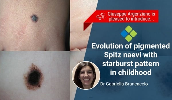
Dr Gabriella Brancaccio provides a new dermoscopy update on the evolution of pigmented Spitz naevi with starburst pattern during childhood.
-1.jpg)
This flat 8x9mm pigmented lesion is found on the back of a 45-year-old male who presents for a follow-up skin check following a melanoma diagnosis.
.jpg)
Over 90 per cent of skin cancers are managed by GPs. Learn from Dr Tony Azzi about the importance of GPs providing skin cancer treatments in primary care.
-1.jpg)
Case discussion: What is your differential diagnosis for this painless, non-healing ulcer on the foot of an 85-year-old male patient?
.jpg)
In this HealthCert podcast, Paul Elmslie speaks to Dr Terry Harvey about his journey as a skin cancer doctor practising on the Sunshine Coast.