1800 867 1390
How would you manage this lesion on the back of a female patient with a history of melanoma which total body photography AI skin imaging identifies as new?
HealthCert Education
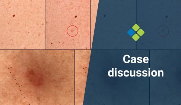
This week we revisit a case from Dr Terry Harvey's practice. A female patient presents for a follow-up skin check in the setting of a previous melanoma. She has total body photography repeated with each of her skin checks.
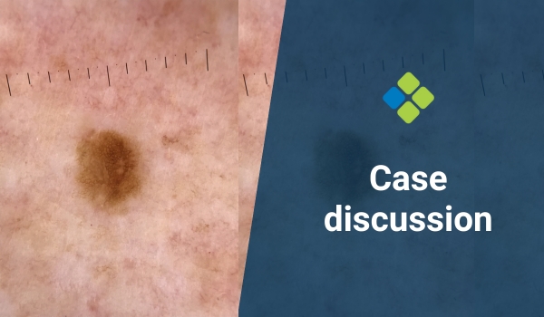
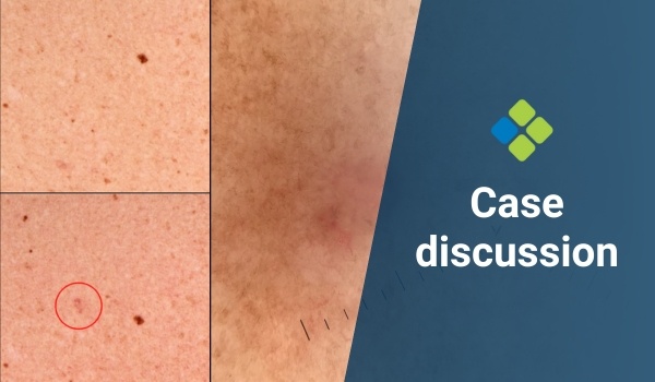
Below is a comparison of her previous imaging (left) and her latest imaging (right). The artificial intelligence identifies the lesion circled in red as new from her previous images.
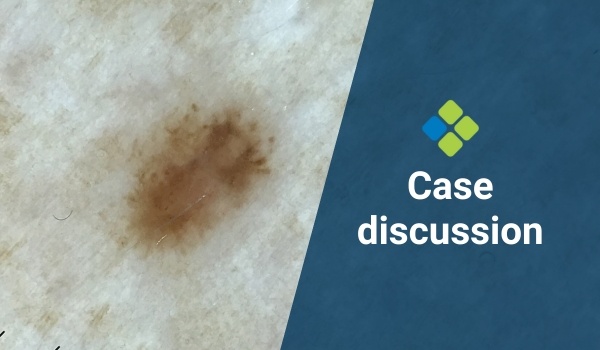
The dermoscopy photo is also of the circled lesion, which is located on her back.
Please refer to the total body photography and dermoscopy images below. What would you do here?


Update
The lesion was excised, and the pathology returned as nodular basal cell carcinoma with clear margins.

 |
Participate with your cases so that we can learn together! Submit your case here or send details to admin@healthcert.com |
| Contributing to the Skin Cancer Case Discussion Blog helps meet your annual Performance Review CPD requirement! |
|
| Submit your own case* = 1 CPD hour (Performance Review) *Case must be published on the blog to qualify. |
Comment/engage with colleagues’ cases = 0.5 CPD hours (Performance Review) |
 How to claim your CPD hours How to claim your CPD hoursIf you interact with this case or submit your own case, you can Quick Log your CPD hours with the RACGP via the usual self-submission process. You will be asked to reflect on what you have learned, and you will require proof you interacted with the blog; a screenshot will suffice. |
|