
 1800 867 1390
How would you manage this 56-year-old man who presents for his annual skin check after his wife comments on this standout, black lesion on his back?
-1.jpg)
HealthCert Education
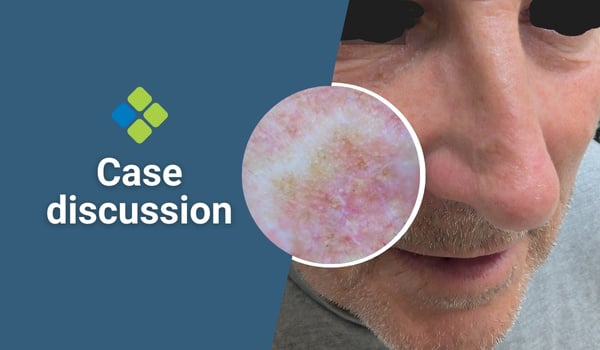
In this week's case discussion, from Dr John O'Bryen, we look at a 56-year-old man who presents for his annual skin check. His wife comments on this standout, black lesion on his back.
The dermoscopy photo is taken in non-contact, polarised mode.
What is your diagnosis, and what would you do next?


Update
A shave removal of the lesion reports seborrhoeic keratosis.
A contact, non-polarised dermoscopy may have shown white clods and helped support the diagnosis. However, sometimes it is better to remove and test lesions like this to reassure our patients. If I was suspicious for melanoma I would have done an excisional biopsy. - Dr John O'Bryen

 |
Participate with your cases so that we can learn together! Submit your case here or send details to admin@healthcert.com |
| Contributing to the Skin Cancer Case Discussion Blog helps meet your annual Performance Review CPD requirement! |
|
| Submit your own case* = 1 CPD hour (Performance Review) *Case must be published on the blog to qualify. |
Comment/engage with colleagues’ cases = 0.5 CPD hours (Performance Review) |
 How to claim your CPD hours How to claim your CPD hoursIf you interact with this case or submit your own case, you can Quick Log your CPD hours with the RACGP via the usual self-submission process. You will be asked to reflect on what you have learned, and you will require proof you interacted with the blog; a screenshot will suffice. |
|
.jpg)
.jpg)


.png?width=100&height=100&name=Jules%20Wynyard%20edit%20circle%20(3).png)