In this webinar and Q&A, solicitor and former regulator David Gardner provides exclusive insights...

Diagnose difficult benign lesions, rare skin tumours, melanoma, BCC, and keratinocyte skin cancers.
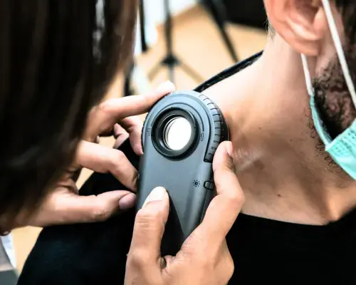
With teachings from the world’s most influential dermoscopy experts, advance your knowledge in the management of nail lesions, mucosal lesions, difficult benign lesions, melanomas, pink tumours, rare skin tumours, BCC, and more.
- Ideal for practitioners who already use dermoscopy and are interested in advancing their skill-set.
- Delivered in collaboration with the International Dermoscopy Society and CPD-accredited.
- Presented by the world's leading dermoscopy experts including Prof Harald Kittler (Austria), Prof Ashfaq A. Marghoob (USA), A/Prof Iris Zalaudek (Austria), Prof Luc Thomas (France), A/Prof Andreas Blum (Germany), A/Prof Aimilios Lallas (Greece), and more.
- This course is for medical doctors, International Medical Graduates, registered nurses, and degree-qualified health professionals.
Fulfils 50 hrs for medical professionals in Australia*
100% online
$2095
Special rates available
80 hrs
Self-paced
*provided an outcome measurement activity with a minimum of 5 hours is completed.

- Improve your accuracy in identifying a broad range of skin lesions, including pink tumours, mucosal lesions, and difficult benign lesions.
- Expertly evaluate nail lesions and detect potential issues early when chances for cure are best.
- Learn to detect and manage rare skin tumours to ensure you provide the most comprehensive care.
- Understand the broad spectrum of BCC and keratinocyte skin cancers for timely detection.
- Learn how to use dermoscopy for general dermatology cases, improving overall patient care.
This module focuses on the general aspects of the dermoscopic examination of pink tumours. It explains the patterns associated with melanocytic and non-melanocytic skins tumours in detail. This module also explains the contact and non-contact dermoscopy methods used when examining skin tumours. Dermoscopic images are used throughout the presentation to identify if the pink tumours are benign or suspicious based on vascular structures, arrangements, and clues. The module concludes with management rules to follow in order not to miss amelanotic melanoma.
This module introduces the basics of mucosal areas including tissue structure and then discusses a variety of mucosal lesions. It explains the clues to use during the clinical and dermoscopic examination together with supporting images. The module then focuses on dermoscopic features of malignant mucosal lesions that are best to be diagnosed in the early stages. It also explains the dermoscopic features of a benign mucosal lesion to avoid any unnecessary excisions. Dermoscopic images are used to evaluate the dermoscopic patterns of mucosal lesions and to identify if a lesion is benign or malignant.
This module discusses the use of dermoscopic clues and features in diagnosing benign lesions that are often challenging to recognise. The module explains these clues and features in detail including imaging, to improve the recognition of melanocytic and non-melanocytic benign tumours that might stimulate melanoma. The module also outlines the four main simulator scenarios; melanoma-like nevi, melanoma-like seborrheic keratosis, spitzoid-looking lesions, and nevi with special features.
This module focuses on the diagnosis of nail pigmentation with dermoscopy. Dermoscopic images of nail pigmentation are used throughout the presentation to establish differential diagnoses between melanoma and other conditions. The module explains the use of dermoscopic clues, patterns and clinical algorithms to diagnose melanonychia striata longitudinal and to determine if a biopsy is needed. The module also discusses the importance of diagnosing the conditions in the early stages and providing conservative treatment to reduce the likelihood of disability.
This module focuses on dermatoscopic clues whilst diagnosing and managing rare, complex syndrome skin tumours. A multidisciplinary approach is the key to managing, diagnosing and treating a rare skin tumour. The module discussed the various types of rare skin tumours such as tumours of fibrous tissue, Merkel cell carcinoma, angiosarcoma, adnexal tumours and sebaceous tumours with the help of dermoscopic images. The module also explains in detail, the dermoscopic clues, patterns, and management of each of these rare skin tumours.
This module discusses the broad spectrum of BCC and keratinocyte skin cancer such as actinic keratosis, Bowen’s disease, intraepithelial carcinoma as well as keratoacanthoma and invasive SCC. The module explains the dermoscopic clues, clichés and common pitfalls whilst diagnosing a BCC. It also talks about the rare and important subtypes of BCC. Extensive images and examples are provided to identify the different types of skin cancer.
This module talks about the dermoscopic clues used to identify melanomas that are difficult to diagnose clinically because of their morphology. It highlights the use of dermoscopy to improve the pattern recognition of various types of melanomas including nodular, desmoplastic, amelanotic, epidermotropic metastatic, nevoid, verrucous and melanomas on sun-damaged skin. The dermoscopic images, features and methods to safely deal with these melanomas are discussed in detail throughout this module.
This module focuses on the significance of integrating dermoscopy in the basic practice of general dermatology. In this module, common dermatological conditions including psoriasis, dermatitis, lichen planus, pityriasis rosacea, acne, dermatitis or fungoides are discussed in detail. It also explains using clinical examinations, laboratory investigations and imaging examinations to achieve an accurate diagnosis. There are parameters and criteria such as vessels morphology, vessel distribution, scale colour, scale distribution, follicular disturbances and specific clues that need to be evaluated before reaching a diagnosis. Dermoscopic images are used throughout the presentation to discuss these parameters and to assist in differentiating between inflammatory and infectious skin diseases.
If you're not interested in pursuing a full certificate in this field but simply want to enhance your skills in specific topics covered in this course, you can access the content of this and other courses for a flat fee of $83 per month (paid annually) within HealthCert 365.
Professor at the Department of Dermatology, Medical University of Vienna, Austria
Professor Harald Kittler has a special clinical interest in dermoscopy of pigmented skin lesions. His main research interest is digital dermoscopy, follow-up of pigmented skin lesions, and computer assisted digital dermoscopy. Harald has been working for 10 years in the field of dermoscopy and has published a number of scientific articles especially in the field of digital dermoscopy and dermoscopic follow-up of melanocytic nevi.
Attending Physician, Dermatology Service, Memorial Sloan Kettering Skin Cancer Center, New York, USA
Professor Ashfaq A. Marghoob is a board-certified dermatologist specialising in the diagnosis and treatment of cancers of the skin. He is the director of Memorial Sloan Kettering’s regional skin cancer clinic in Long Island and consults and treats patients in the centre’s outpatient facility in Manhattan.
Although providing the best care possible for his patients remains his primary goal, Ashfaq also remains committed to education and clinical research, with the hope of educating physicians and the public about the importance of early skin cancer detection to save lives.
He is active in clinical research and has published numerous papers on topics related to skin cancer with an emphasis on melanoma, atypical/dysplastic nevi, and congenital melanocytic nevi. Ashfaq’s research interests are focused on the use of imaging instruments such as photography, dermoscopy, and confocal laser microscopy to recognise skin cancer early in its development.
Professor and Chairman Department of Dermatology, Medical University of Lyon, France
Professor Luc Thomas was board-certified in dermatology in 1989 at Lyon 1 University. He was trained as a post-doctoral fellow at Harvard Medical School in 1990 and 1991, and obtained his PhD degree at Lyon 1 University in 1993. He became full professor of dermatology in 1996, first class professor in dermatology in 2009, and Chairman of the Department of Dermatology of Lyon 1 University - Centre Hospitalier Lyon Sud in 2003. He obtained his Board certification in Clinical Oncology in 2013.
Luc’s main research fields include skin oncology, early diagnosis of melanoma, dermoscopy, skin surgery and nail diseases. He has published more than 400 peer-reviewed scientific articles in international journals, is the co-editor of four books published in several languages and co-author of more than 25 books. He has lectured at many international meetings, is an associate editor of Dermatology, a member of the board of the International Dermoscopy Society, a past member of the board and treasurer of the French Society of Dermatology from 2000 to 2003, and treasurer of the World Congress of Dermatology in Paris in 2002.

Public, Private and Teaching Practice, Konstanz, Germany
Associate Professor at the University of Tübingen, Germany
Associate Professor Andreas Blum studied medicine in Germany and France and received his Doctor of Medicine degree in 1993. From 1993 until 2004, he worked in the Department of Dermatology at the University of Tübingen in Germany. In 1998 he finished the specialisation in Dermatology and Venereology. Since 2002 he has been a senior lecture and assistant professor and, since 2006, has been an associate professor at the University of Tübingen. Since 2004 he has worked in his private and teaching practice in Konstanz, Germany.
Andreas is an expert in the diagnosis, surgical treatment, prevention and follow-up of skin cancers. In addition to his clinical research mainly in the field of dermoscopy, he gives regular lectures for national and international dermatological societies.

Dermatologist-Venereologist, First Department Of Dermatology, Aristotle University, Greece
President, International Dermoscopy Society
Aimilios Lallas is an Associate Professor of Dermatology at the First Department of Dermatology of Aristotle University in Thessaloniki, Greece. He is specialised in skin cancer diagnosis with non-invasive techniques, as well as in the management of skin cancer patients.
His main field of research interest is dermoscopy of skin tumours, the application of the method in general dermatology and the improvement of the management of oncologic patients. He is an author of more than 330 scientific papers published on Pubmed Central, most of them on dermoscopy and skin cancer. He is an editor of eight books and author of several book chapters on dermoscopy. He is a co-investigator in several Phase III Clinical trials on skin cancer treatment. He has been awarded several scholarships and scientific awards.
Over the last years, A/Prof Lallas has established scientific collaboration with numerous colleagues from several countries and has supervised the training of numerous fellows from different countries. He is an invited speaker in several domestic and international congresses and meetings, mainly on dermoscopy and on skin cancer diagnosis and management. He is particularly involved in teaching activities on dermoscopy, having organised and participated in numerous domestic and international courses.
A/Prof Lallas is currently the President of the International Dermoscopy Society.

Scientific Coordinator, Skin Cancer Unit, ASMN-IRCCS, University of Modena and Reggio Emilia, Italy
Associate Professor Caterina Longo is a board-certified dermatologist specialising in the diagnosis and treatment of skin cancers. Although providing the best care possible for patients remains her primary goal, she also committed to education and clinical research. She is actively involved in clinical research and has published numerous papers on topics related to skin cancer with an emphasis on melanoma, atypical nevi, Spitz/Reed nevi and non-melanoma skin cancer.
Caterina’s research interests are focused on the use of imaging instruments such as dermoscopy and confocal laser microscopy to recognise skin cancer early in its development. She pioneered the use of ex vivo fluorescence confocal microscopy for micrographic Mohs surgery applied for basal cell carcinoma and other visceral tumours. Caterina lectures on these topics both nationally and internationally.

Head of the Dermatology Clinic of the University of Trieste, Italy
Associate Professor Iris Zalaudek is a board-certified dermatologist and Head of the Dermatology Clinic of the University of Trieste, Italy. Since 2016, she has been President of the International Dermoscopy Society, and was previously the Research Director of the Non-Melanoma Skin Cancer Unit at the Medical University of Graz, Austria.
Her main research fields are related to dermato-oncology and include non-invasive skin imaging techniques, as well as topical and systemic treatment of skin cancer. Moreover, she is engaged in the development of modern teaching methods such as online distant courses and tele-dermatologic services. She is Director of the Master of Science program entitled "Dermoscopy and Preventive Dermato-Oncology" of the Medical University of Graz, Austria.
Iris has published more than 450 articles, of which 358 (267 full papers) have been cited in PubMed. Her combined publications have received an impact factor of 1003 and a h-index value of 36 (by April 2017). In 2003 her work was awarded by the Hans-Weitgasser Price from the Styrian Association of Dermatologists and in 2008 she was awarded the Best Researcher of the Medical University of Graz, Austria.

Dermatologist, Santa Maria Nuova Hospital, Reggio Emilia, Italy
Dr Elvira Moscarella is a dermatologist at the Santa Maria Nuova Hospital in Reggio Emilia, Italy. She acquired her medical degree in 2005 at the Second University of Naples before completing her residency in dermatology and venereology at the University’s Department of Dermatology. In 2008, Elvira undertook further education in dermoscopy and confocal microscopy. She is a member of the European Academy of Dermatology and Venereology and the International Dermoscopy Society, and is Editor in Chief of the latter’s newsletter and case of the month. Elvira’s main interests are in dermoscopy and reflectance confocal microscopy, and their use in skin cancer medicine.

Study at your own pace and to your own schedule. Interactivity, discussion, and feedback opportunities are included.

Easily meet your CPD requirements and gain valuable skills – all in one place for $83 per month.
$2095
.
*provided an outcome measurement activity with a minimum of 5 hours is completed.
Bundle two courses and save 5%, or three courses and save 10% upon enrolment.
Talk to us about deferred payment options, registrar scholarships and special rates.

I think the Advanced Certificate of Dermoscopy was a terrific roundup of assistance in difficult areas. It is an efficient and enjoyable way to consolidate and extend your dermoscopy skills. I enjoyed the course and am looking forward to the next stage. The web-based self paced mode of presentation is very helpful as it avoids adding travel time and pet-care challenges to participate compared with physically attending in a major city.
Dr Y Wylde
This is an excellent course for general practitioners. Excellent content, clearly explained. Thank you for giving me a wonderful learning experience.
Dr N. S. K. Liyanage
Excellent quality of content. I love the enthusiasm that comes through in all of the lectures. Thank you for making this an enjoyable learning experience.
Dr M. Vukic
In-depth e-learning on dermoscopy! This is a great course for learning dermoscopy on special sites and general dermatology!
Dr M. Spit
I really enjoyed the level of learning. It is very rewarding to know that I am potentially saving lives. Recently I volunteered with the Lions Cancer Institute for two days, and we screened 158 patients, detected 38 possible melanomas and 83 keratinocyte skin cancers. It was a very successful and rewarding two days, and something I could do confidently because of my learning from this course.
Dr K. Laverty
| RACGP Activity Number | ACRRM Activity Number | Activity Title | Education Hours | Performance Hours | Outcome Hours | ||
|---|---|---|---|---|---|---|---|
| 405086 | 28496 | Difficult benign lesions including nevi with special features | 405086 | 28496 | 4 | 6 | 0 |
| 405101 | 28501 | Dermoscopy in general dermatology | 405101 | 28501 | 4 | 6 | 0 |
| 405076 | 28490 | Mucosal lesions | 405076 | 28490 | 4 | 6 | 0 |
| 405100 | 28500 | Difficult melanomas | 405100 | 28500 | 4 | 6 | 0 |
| 405092 | 28498 | Rare skin tumours | 405092 | 28498 | 4 | 6 | 0 |
| 405099 | 28499 | The broad spectrum of BCC and keratinocyte skin cancer | 405099 | 28499 | 4 | 6 | 0 |
| 405070 | 28489 | Pink tumours | 405070 | 28489 | 4 | 6 | 0 |
| 405089 | 28497 | Nail lesions | 405089 | 28497 | 4 | 6 | 0 |
| 808030 | 32974 | Difficult to Diagnose Melanomas Outcome Improvement Activity | 808030 | 32974 | 0 | 0 | 8.5 |
| Total hours | 32 | 48 | 8.5 | ||||
The purpose of outcome measurement activities is to improve your clinical confidence in managing an identified learning gap. Outcome measurement activities are not a requirement of our Professional Certificate of Advanced Certificate courses; they are a requirement for Australian CPD purposes.
HealthCert Education provides a variety of outcome measurements activities to suit your needs:
The Advanced Certificate of Dermoscopy will meet the needs of medical professionals who already use dermoscopy in their practice and are interested in taking their skill-set to a more advanced level. Participants will acquire in-depth knowledge in lesion management, including difficult and rare lesions, with teachings by an accomplished team of dermatologists and dermoscopy experts.
The course is suitable for medical doctors and the degree-qualified nurses who work under their supervision, other degree-qualified health professionals with an interest in skin, as well as for International Medical Graduates.
Participants must have successfully completed the HealthCert Professional Certificate of Dermoscopy course (or a qualification deemed equivalent) and HealthCert also highly recommends successful completion of at least 25 cases of dermoscopy prior to enrolment. Equivalent alternatives include: practical experience, other qualifications or successful completion of the Professional Certificate examination.
Participants do not have to pass an IELTS test but, as the courses are delivered in English, proficiency in listening, reading and writing English is assumed.
Participants will require access to a computer/laptop, an internet connection and a basic level of technology proficiency to access and navigate the online learning portal.
Professionally accredited qualifications and prior studies may be recognised for entry into this course. Please send an email to credit@healthcert.com for an individual assessment of your prior qualifications and experience. This email should contain information about your educational history and work experience that specifically pertain to the content and procedures covered in the Professional Certificate of Dermoscopy. Please include any applicable certificates and course outlines from previous education. The relevant Course Chair will make a determination on your application within two to three weeks.
Doctors who have completed the UQ Certificate of Advanced Dermatoscopy and Histopathology or other formal dermoscopy training can receive academic credit towards the Professional Diploma (the final course in the three-part program) if they achieve a pass mark in the exams of the first two certificate courses Professional Certificate of Dermoscopy and Advanced Certificate of Dermoscopy. Upon successful exam completion doctors can directly progress to (and will only need to pay for) the Professional Diploma of Dermoscopy course.
NOTE: While the Professional Certificate of Skin Cancer Medicine course covers Dermoscopy, it does not qualify for recognition of prior learning in the Certificate and Professional Diploma of Dermoscopy program which quickly moves on to more advanced dermoscopy techniques, including the Chaos and Clues method and the assessment of lesions on the face and acral sites. Professional Certificate of Skin Cancer Medicine alumni should begin with the Professional Certificate of Dermoscopy course.
This certificate course meets the minimum 50 hours CPD annual requirement across all three mandatory CPD activity types, provided an outcome measurement activity with a minimum of five hours is completed. You may use an optional HealthCert outcome measurement activity or develop your own.
Outcome measurement activities are not a requirement of Professional or Advanced Certificates.
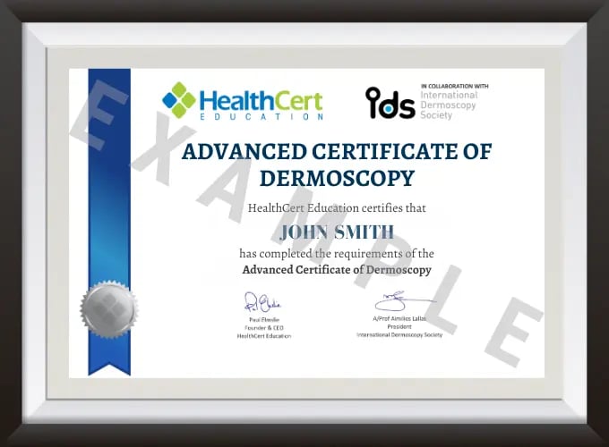
Upon successful completion of the course requirements, course participants will receive the certificate of the Advanced Certificate of Dermoscopy.
This certificate course:
Australia-based course participants, please note that HealthCert certificates are not accredited by TEQSA or ASQA and do not fall within the Australian Qualification Framework. All HealthCert certificates are professional development awards, accredited by RACGP, ACRRM and RNZCGP.
To learn more about the delivery of certificates in Australia and overseas, please visit our FAQs.
Professional Diploma Pathway
This course is the second stage of the three-part professional diploma pathway. The full pathway is Professional Certificate of Dermoscopy, Advanced Certificate of Dermoscopy, and Professional Diploma of Dermoscopy.
RPL with The University of Queensland
The University of Queensland Master of Medicine (Skin Cancer) includes the unit IMED7002: Clinical and Dermatoscopic Diagnosis in Skin Cancer Practice. Credit precedence has been established for this subject if students have completed all three of the following HealthCert dermoscopy qualifications: Professional Certificate of Dermoscopy, Advanced Certificate of Dermoscopy and Professional Diploma of Dermoscopy to the unit IMED7002. Doctors who have applied and been accepted into the Master of Medicine (Skin Cancer) program may apply for Recognition of Prior Learning for IMED7002. View The University of Queensland's Master of Medicine (Skin Cancer) program here.
Master of Medicine (Skin Cancer)
Doctors who complete the HealthCert Professional Diploma programs in Dermoscopy, Skin Cancer Medicine and Skin Cancer Surgery will receive RPL for the units IMED7002, IMED7010, and IMED7011, which are high-level subjects in the Master of Medicine (Skin Cancer) at The University of Queensland. The Master of Medicine is open only to registered medical practitioners with at least two years' postgraduate experience. View The University of Queensland, Master of Medicine (Skin Cancer) program.
Please note, only HealthCert Education qualifications completed within the last ten years can be recognised.
Southern Cross affiliated provider
For medical practitioners in New Zealand, completion of this certificate course enables you to become an affiliated provider with Southern Cross as a Skin Cancer Medicine Doctor.
HealthCert Clinical Attachments
Course participants who successfully complete the HealthCert Professional Certificate of Skin Cancer Medicine may continue their professional development by completing a HealthCert Clinical Attachment at a clinic or university teaching hospital to further develop professional knowledge. A HealthCert Australian Clinical Attachment is recommended as the first clinical attachment after completing the HealthCert dermoscopy qualifications and a HealthCert International Clinical Attachment is recommended for subsequent clinical attachments.
This organisation is an RACGP-accredited CPD provider under the RACGP CPD Program.

Don't see your question? Explore other faqs or talk to us.
Fees will vary based on the program and study option selected (fully online vs online + optional practical workshop). Payments can be made upfront or in monthly instalments. Special rates and various payment options are available. GP registrars and doctors in training enjoy a scholarship of up to $500. Talk to us to learn more.
Completion of any HealthCert course or attendance at an event will enable you to access the HealthCert Alumni Program which includes:
HealthCert Education is pleased to issue digital credentials for alumni. Digital credentials are a permanent online record of your successful completion of a HealthCert course and are issued to all course participants in addition to PDF certificates. If you are based in Australia, you also have the option to order a hard copy of your digital certificate for a small additional fee.
The recommended study duration of this certificate course is 80 hours, which includes study of the pre-course activities and readings, online lectures, live tutorials, and online assessment. This self-paced course offers the flexibility of 100% online study in your own time, at your own pace, in your own home or office, with no mandatory face-to-face requirements. You are not required to be online at specific times but can view and replay video lectures at your convenience.
All HealthCert courses meet World Federation of Medical Education standards. This certificate course qualifies for CPD hours from the Royal Australian College of General Practitioners (RACGP) and the Australian College of Rural and Remote Medicine (ACRRM) in Australia. It is recognised by the Royal New Zealand College of General Practitioners (RNZCGP) in New Zealand. It is recognised by the Hong Kong College of Family Physicians (HKCFP) in China. It is a self-submitted activity in Dubai and the United Kingdom. It is a self-submitted activity through the College of Family Physicians in Canada. If you live or work outside one of the above-mentioned countries, please contact us on admin@healthcert.com to discuss whether this course can be recognised in your country.

In this webinar and Q&A, solicitor and former regulator David Gardner provides exclusive insights...
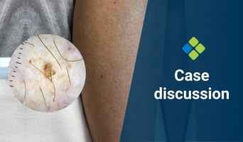
This week's case discussion, submitted by Dr Heather Lawson, features a 64-year-old female patient...

Mood changes in the postpartum period are extremely common, ranging from mild and transient “baby...
Want to stay up-to-date with the latest case studies, podcasts, free video tutorials and medical research articles pertinent to primary care?
Our Education Advisors can assist you with any queries and tailor our education pathway to suit your current expertise, interests and career goals.